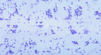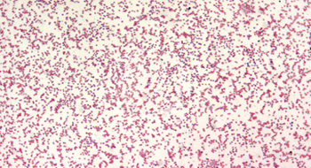

Tularemia is caused by the most infectious pathogenic bacteria known to man, Francisella tularensis. It is a zoonotic disease that can cause a severe and prolonged nonspecific febrile (fever) illness lasting from several weeks to months. This disease can be acquired in a variety of ways, but most commonly through bites from arthropods, such as ticks, biting flies, and mosquitoes. It can also be transmitted through inhalation, direct contact, or ingestion of contaminated animal tissue (often rabbits and rodents), soil, or water. F. tularensis is found throughout rural areas in North America and Eurasia. In the United States, approximately 126 human cases are reported each year, with less than 2% being fatal. Most cases occur in rural areas of the south-central and western states during summer months. From 2000 to 2008, 57% of all reported tularemia cases in the U.S. were located in Missouri, Arkansas, Oklahoma, Massachusetts and South Dakota. There are two main subspecies of F. tularensis, subspecies tularensis (Type A), the dominant strain North America, and subspecies holarctica (Type B), the only disease causing strain in Eurasia. Type A is highly virulent, whereas Type B produces milder forms of the disease and is rarely fatal.
F. tularensis is considered an effective bioweapon because it can be aerosolized, it has an extremely low infectious dose (~10 organisms), and can cause a substantial, long-lasting illness, sometimes leading to death. Although the bacteria does not form spores, F. tularensis can survive for weeks at low temperatures in moist soil, water, hay, straw, or decaying plants and animals. Although F. tularensis has a low mortality rate, an aerosol delivery over a metropolitan area could incapacitate hundreds of thousands of people for weeks to months, draining valuable medical resources. It has been studied by the former biological weapons programs of Japan, the United States, which produced less than 2 tons per year, and the Soviet Union, which produced over one thousand tons of F. tularensis per year.
The virulence factors for F. tularensis are largely unknown, but are currently under investigation. Once F. tularensis is engulfed by immune cells (macrophages) that routinely kill bacteria, it replicates and evades parts of the immune system. A few proteins known to be involved in this process include MglA, IglC, AcpA and MinD. MglA regulates the activity of the F. tularensis pathogenicity island, a portion of the genome with multiple virulence genes. One of these genes codes for the protein Iglc, which is thought to disrupt a component of immune system that can recognize the presence of bacteria (TLR-4 signaling). Without this recognition, the immune response against a bacterial infection will be less effective. The AcpA and MinD proteins help protect F. tularensis from oxidative bursts, a collection of cell-damaging free radicals released by immune cells to kill bacteria. F. tularensis also infects the lymph nodes, lungs, spleen, liver, and kidneys. Continued bacterial growth in these areas leads to lesion formation on and within the infected organs, inflammation and necrosis of surrounding tissue.
Tularemia symptoms usually appear within 3-5 days of infection, but may also appear up to 15 days later. Initial symptoms include fever, chills, headaches, diarrhea, muscle aches, joint pain, dry cough, sore throat, and progressive weakness. Prolonged infection can result in pneumonia, chest pain, bloody sputum, weight loss, difficulty breathing, and death. If untreated, the disease can last for months. Depending on the route of infection and the disease presentation, tularemia can be classified as glandular, ulceroglandular, oculoglandular, oropharyngeal, typhoidal, pneumonic, and/or septic; many symptoms overlap between these classifications. Glandular tularemia presents with swollen lymph nodes. Ulceroglandular tularemia is characterized by a skin ulcer (sore) at the site of infection and swollen lymph nodes. Oculoglandular tularemia results from the entrance of F. tularensis through the eye, and is characterized by painful purulent conjunctivitis (red, irritated eye with a discharge of pus) and swollen lymph nodes. Oropharyngeal tularemia results from ingesting contaminated food or water and is characterized by severe exudative pharyngitis (oozing sore throat). Typhoidal tularemia presents with the standard, nonspecific febrile (fever) symptoms mentioned earlier, but without a known route of infection (via skin, eye, ingestion or inhalation). Direct inhalation of aerosolized F. tularensis or secondary spread of the bacteria can lead to pneumonic tularemia, an infection of the lungs. Approximately 30% of patients with ulceroglandular tularemia, and 80% with typhoidal tularemia, will develop pneumonic tularemia. Untreated tularemia may progress into septic tularemia, where the body goes into a state of septic shock due to an overwhelming bacterial infection. Untreated tularemia caused by the more virulent tularensis (Type A) subspecies has a reported overall mortality of 5-15%, which increases to 30-60% for pneumonic and septic forms.
Suspected cases oF. tularemia are treated with antibiotics, including streptomycin, gentamicin, doxycycline, and ciprofloxacin. Preliminary testing for a tularemia infection can be done in two hours; however, confirmation can take another 24-48 hours. Mortality is less than 2% with treatment.
The tularemia vaccine, the Live Vaccine Strain (LVS) of F. tularensis, was originally developed in the Soviet Union and transferred to the United States in 1956. This vaccine is classified as an investigational new drug (IND) by the Food and Drug Administration (FDA), and is only available to laboratory workers and others at high-risk for infection. This vaccine can fully protect against low-dose aerosols and partially protect against high-dose aerosols of F. tularensis.
Dembek, Z.F. [Senior Editor] (2007). Tularemia, Chapter 8, Medical Aspects of Chemical and Biological Warfare. Publisher: Department of Defense, Office of The Surgeon General, US Army, Borden Institute. 2007: 672 p.; ill http://www.bordeninstitute.army.mil/published_volumes/biological_warfare/biological.html
Dennis, D.T, et al. (2001). Tularemia as a Biological Weapon. JAMA, Vol. 285, No. 21: 2763-2773.
Centers for Disease Control, USA (2009). Reported tularemia cases by state -- United States, 2000 – 2008. Accessed 06 May 2010 from http://www.cdc.gov/tularemia/Surveillance/Tul_CasesbyState.html
Berrada Z.L. et al. (2006). Raccoons and Skunks as Sentinels for Enzootic Tularemia. Emerging Infectious Diseases, Vol. 12, No. 6: 1019-1021
Feldman, K.A., et al. (2003). Tularemia on Martha’s Vineyard: seroprevalence and occupational risk. Emerging Infectious Diseases, Vol. 9, No.3:350–354.
Croddy, E. & Krcálová, S. (2001). Tularemia, Biological Warfare, and the Battle for Stalingrad (1942-1943). Military Medicine, Vol. 166, No. 10: 837-8838. Accessed 06 May 2010 from http://cns.miis.edu/archive/cbw/tula.htm